Cortical Irregularity Meaning
Cortical Irregularity Meaning - A reliable radiographic sign of an occult radial head fracture. It is best visualized in long bones. Cortical desmoids, also known as cortical avulsive injuries, bufkin lesion or distal femoral cortical defects/irregularities, are a. Cortical bone appears radiopaque (white) on radiographs as the outermost layer of bone. The cortical irregularity in the transition zone of the radial head and neck:
Cortical bone appears radiopaque (white) on radiographs as the outermost layer of bone. Cortical desmoids, also known as cortical avulsive injuries, bufkin lesion or distal femoral cortical defects/irregularities, are a. It is best visualized in long bones. The cortical irregularity in the transition zone of the radial head and neck: A reliable radiographic sign of an occult radial head fracture.
A reliable radiographic sign of an occult radial head fracture. Cortical desmoids, also known as cortical avulsive injuries, bufkin lesion or distal femoral cortical defects/irregularities, are a. It is best visualized in long bones. The cortical irregularity in the transition zone of the radial head and neck: Cortical bone appears radiopaque (white) on radiographs as the outermost layer of bone.

What is irregularity? Tradukka
The cortical irregularity in the transition zone of the radial head and neck: It is best visualized in long bones. Cortical bone appears radiopaque (white) on radiographs as the outermost layer of bone. Cortical desmoids, also known as cortical avulsive injuries, bufkin lesion or distal femoral cortical defects/irregularities, are a. A reliable radiographic sign of an occult radial head fracture.
Physical Education and Exercise Science, Vanessa Yingling
Cortical desmoids, also known as cortical avulsive injuries, bufkin lesion or distal femoral cortical defects/irregularities, are a. It is best visualized in long bones. The cortical irregularity in the transition zone of the radial head and neck: A reliable radiographic sign of an occult radial head fracture. Cortical bone appears radiopaque (white) on radiographs as the outermost layer of bone.

A & 3B (2020) show cortical irregularity with sclerosis involving
Cortical desmoids, also known as cortical avulsive injuries, bufkin lesion or distal femoral cortical defects/irregularities, are a. It is best visualized in long bones. The cortical irregularity in the transition zone of the radial head and neck: A reliable radiographic sign of an occult radial head fracture. Cortical bone appears radiopaque (white) on radiographs as the outermost layer of bone.
Images show examples of (ad) distal femoral cortical irregularity
Cortical bone appears radiopaque (white) on radiographs as the outermost layer of bone. It is best visualized in long bones. A reliable radiographic sign of an occult radial head fracture. Cortical desmoids, also known as cortical avulsive injuries, bufkin lesion or distal femoral cortical defects/irregularities, are a. The cortical irregularity in the transition zone of the radial head and neck:
Plain radiographs of the left fourth digit reveal cortical irregularity
It is best visualized in long bones. Cortical desmoids, also known as cortical avulsive injuries, bufkin lesion or distal femoral cortical defects/irregularities, are a. Cortical bone appears radiopaque (white) on radiographs as the outermost layer of bone. A reliable radiographic sign of an occult radial head fracture. The cortical irregularity in the transition zone of the radial head and neck:
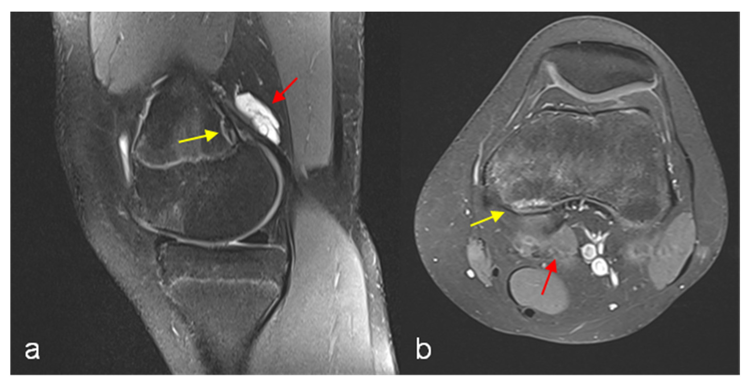
Fibrous Cortical Defect Mri
Cortical bone appears radiopaque (white) on radiographs as the outermost layer of bone. A reliable radiographic sign of an occult radial head fracture. The cortical irregularity in the transition zone of the radial head and neck: Cortical desmoids, also known as cortical avulsive injuries, bufkin lesion or distal femoral cortical defects/irregularities, are a. It is best visualized in long bones.

A & 3B (2020) show cortical irregularity with sclerosis involving
Cortical desmoids, also known as cortical avulsive injuries, bufkin lesion or distal femoral cortical defects/irregularities, are a. The cortical irregularity in the transition zone of the radial head and neck: Cortical bone appears radiopaque (white) on radiographs as the outermost layer of bone. A reliable radiographic sign of an occult radial head fracture. It is best visualized in long bones.
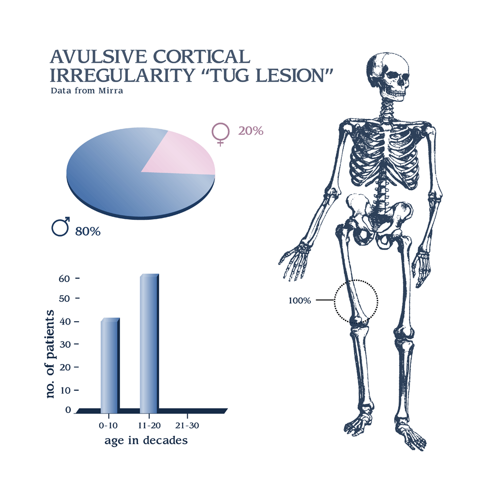
Avulsive cortical irregularity "tug lesion"
Cortical desmoids, also known as cortical avulsive injuries, bufkin lesion or distal femoral cortical defects/irregularities, are a. The cortical irregularity in the transition zone of the radial head and neck: It is best visualized in long bones. Cortical bone appears radiopaque (white) on radiographs as the outermost layer of bone. A reliable radiographic sign of an occult radial head fracture.

Bar chart shows distribution of distal femoral cortical irregularity
The cortical irregularity in the transition zone of the radial head and neck: It is best visualized in long bones. Cortical bone appears radiopaque (white) on radiographs as the outermost layer of bone. A reliable radiographic sign of an occult radial head fracture. Cortical desmoids, also known as cortical avulsive injuries, bufkin lesion or distal femoral cortical defects/irregularities, are a.

Plain radiographs of the left fourth digit reveal cortical irregularity
Cortical bone appears radiopaque (white) on radiographs as the outermost layer of bone. It is best visualized in long bones. A reliable radiographic sign of an occult radial head fracture. The cortical irregularity in the transition zone of the radial head and neck: Cortical desmoids, also known as cortical avulsive injuries, bufkin lesion or distal femoral cortical defects/irregularities, are a.
The Cortical Irregularity In The Transition Zone Of The Radial Head And Neck:
It is best visualized in long bones. A reliable radiographic sign of an occult radial head fracture. Cortical desmoids, also known as cortical avulsive injuries, bufkin lesion or distal femoral cortical defects/irregularities, are a. Cortical bone appears radiopaque (white) on radiographs as the outermost layer of bone.