Inferior Vena Cava
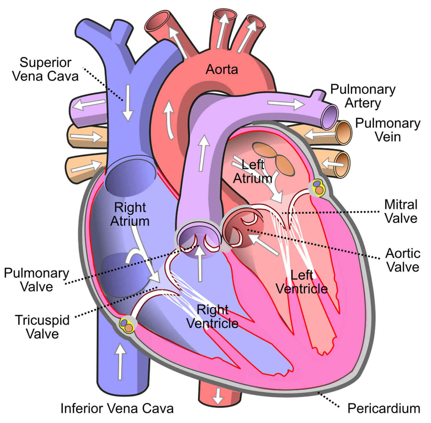
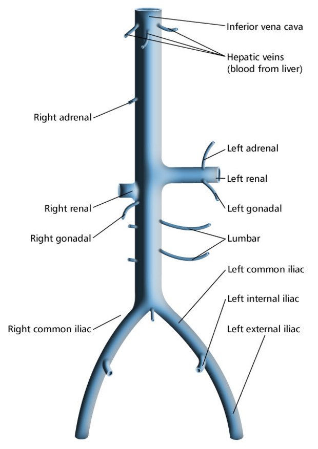
Inferior Vena Cava - It is formed by the joining of the right and the left common iliac veins , usually at the level of the fifth lumbar vertebra. The inferior vena cava is a large vein that carries the deoxygenated blood from the lower and middle body into the right atrium of the heart. The ivc’s function is to carry the venous blood from the lower limbs and abdominopelvic region. The superior vena cava and inferior vena cava are very large veins that bring deoxygenated blood to your heart to get oxygen. Anatomically this usually occurs at the l5 vertebral level. The inferior vena cava (ivc) is the largest vein of the human body. It is located at the posterior abdominal wall on the right side of the aorta. The inferior vena cava (ivc) is a large retroperitoneal vessel formed by the confluence of the right and left common iliac veins.
The inferior vena cava (ivc) is the largest vein of the human body. It is located at the posterior abdominal wall on the right side of the aorta. The inferior vena cava is a large vein that carries the deoxygenated blood from the lower and middle body into the right atrium of the heart. The ivc’s function is to carry the venous blood from the lower limbs and abdominopelvic region. The superior vena cava and inferior vena cava are very large veins that bring deoxygenated blood to your heart to get oxygen. It is formed by the joining of the right and the left common iliac veins , usually at the level of the fifth lumbar vertebra. Anatomically this usually occurs at the l5 vertebral level. The inferior vena cava (ivc) is a large retroperitoneal vessel formed by the confluence of the right and left common iliac veins.
The superior vena cava and inferior vena cava are very large veins that bring deoxygenated blood to your heart to get oxygen. It is formed by the joining of the right and the left common iliac veins , usually at the level of the fifth lumbar vertebra. It is located at the posterior abdominal wall on the right side of the aorta. The inferior vena cava (ivc) is a large retroperitoneal vessel formed by the confluence of the right and left common iliac veins. The inferior vena cava is a large vein that carries the deoxygenated blood from the lower and middle body into the right atrium of the heart. The ivc’s function is to carry the venous blood from the lower limbs and abdominopelvic region. The inferior vena cava (ivc) is the largest vein of the human body. Anatomically this usually occurs at the l5 vertebral level.

Difference Between Superior and Inferior Vena Cava
Anatomically this usually occurs at the l5 vertebral level. It is located at the posterior abdominal wall on the right side of the aorta. The inferior vena cava (ivc) is a large retroperitoneal vessel formed by the confluence of the right and left common iliac veins. The inferior vena cava (ivc) is the largest vein of the human body. The.
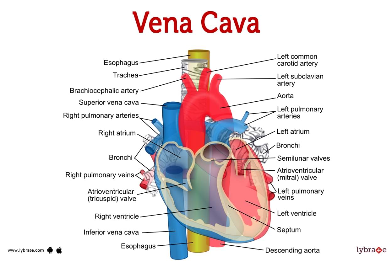
Vena cava (Human Anatomy) Image, Functions, Diseases and Treatments
The inferior vena cava (ivc) is the largest vein of the human body. The superior vena cava and inferior vena cava are very large veins that bring deoxygenated blood to your heart to get oxygen. The inferior vena cava (ivc) is a large retroperitoneal vessel formed by the confluence of the right and left common iliac veins. Anatomically this usually.
inferior vena cava agenesis
The ivc’s function is to carry the venous blood from the lower limbs and abdominopelvic region. The inferior vena cava (ivc) is a large retroperitoneal vessel formed by the confluence of the right and left common iliac veins. The superior vena cava and inferior vena cava are very large veins that bring deoxygenated blood to your heart to get oxygen..
/GettyImages-184897753-ce6892a3766a4256a6951f22ef340269.jpg)
Superior and Inferior Venae Cavae
Anatomically this usually occurs at the l5 vertebral level. It is formed by the joining of the right and the left common iliac veins , usually at the level of the fifth lumbar vertebra. It is located at the posterior abdominal wall on the right side of the aorta. The ivc’s function is to carry the venous blood from the.
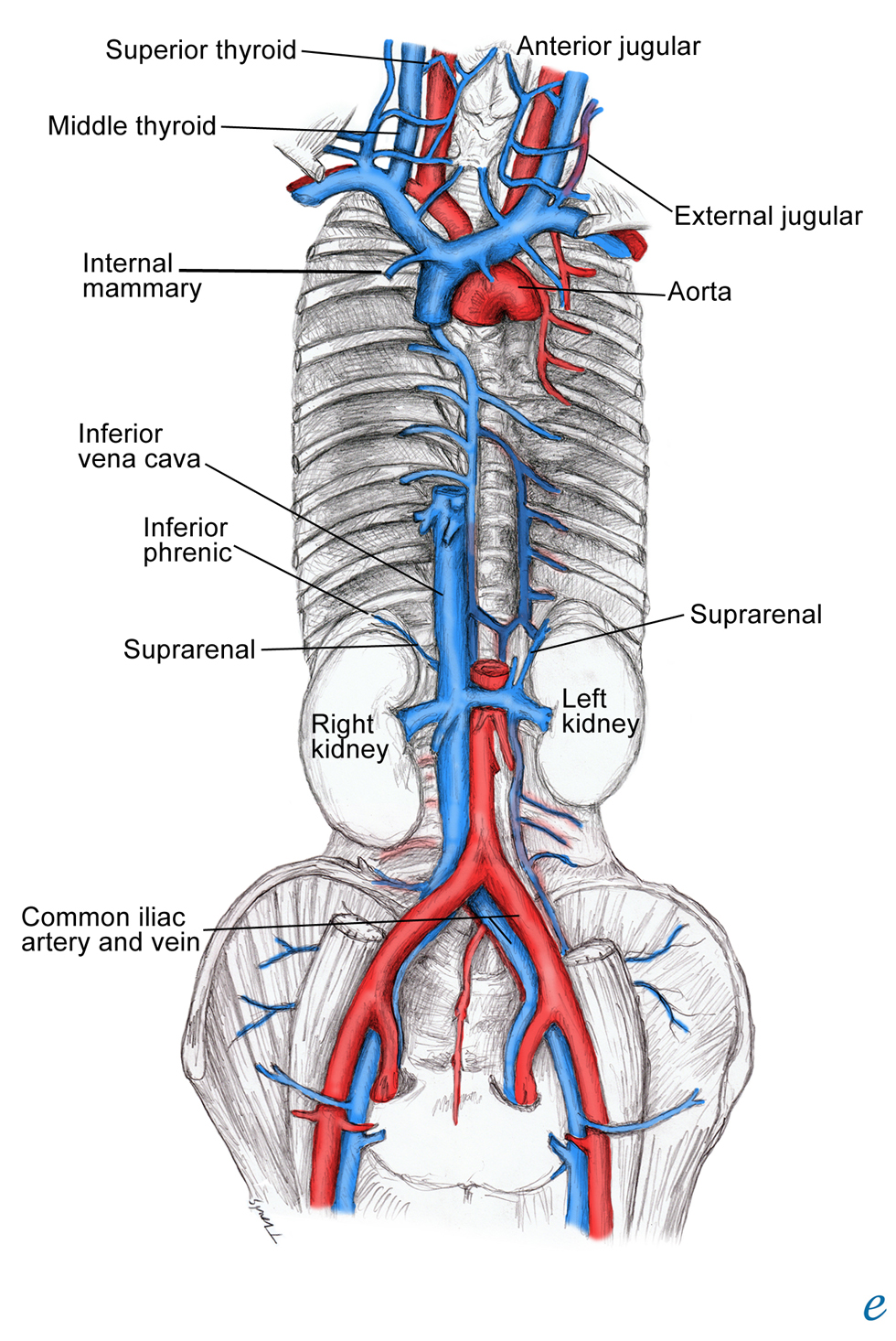
inferior vena cava location
The ivc’s function is to carry the venous blood from the lower limbs and abdominopelvic region. The inferior vena cava is a large vein that carries the deoxygenated blood from the lower and middle body into the right atrium of the heart. It is located at the posterior abdominal wall on the right side of the aorta. The inferior vena.
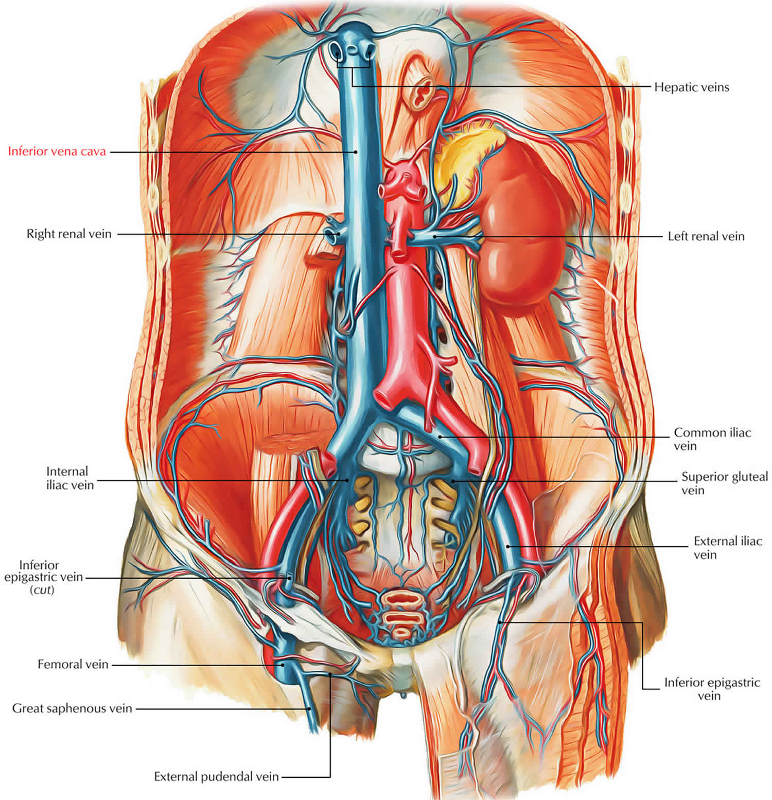
Inferior vena cava anatomy, function, filter & inferior vena cava syndrome
The ivc’s function is to carry the venous blood from the lower limbs and abdominopelvic region. The inferior vena cava (ivc) is a large retroperitoneal vessel formed by the confluence of the right and left common iliac veins. Anatomically this usually occurs at the l5 vertebral level. The superior vena cava and inferior vena cava are very large veins that.

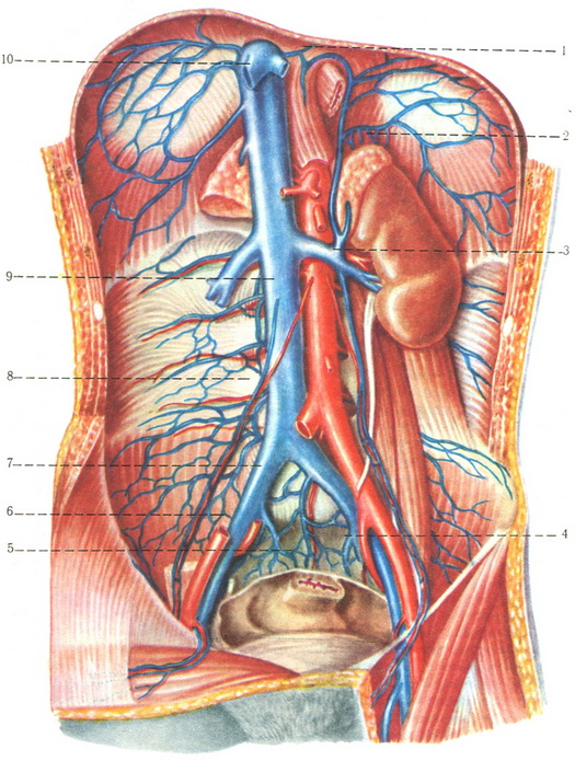
Inferior Vena Cava Overview, Structure & Clinical Significance » How
Anatomically this usually occurs at the l5 vertebral level. The inferior vena cava (ivc) is the largest vein of the human body. The inferior vena cava (ivc) is a large retroperitoneal vessel formed by the confluence of the right and left common iliac veins. It is located at the posterior abdominal wall on the right side of the aorta. The.

Sistema de la vena Cava Inferior, Nombre NOEMY KARLA FERNANDEZ PATON
The inferior vena cava is a large vein that carries the deoxygenated blood from the lower and middle body into the right atrium of the heart. The superior vena cava and inferior vena cava are very large veins that bring deoxygenated blood to your heart to get oxygen. The ivc’s function is to carry the venous blood from the lower.
Inferior vena cava anatomy, function, filter & inferior vena cava syndrome
The inferior vena cava is a large vein that carries the deoxygenated blood from the lower and middle body into the right atrium of the heart. It is located at the posterior abdominal wall on the right side of the aorta. The inferior vena cava (ivc) is the largest vein of the human body. The superior vena cava and inferior.
inferior vena cava syndrome
The inferior vena cava is a large vein that carries the deoxygenated blood from the lower and middle body into the right atrium of the heart. It is located at the posterior abdominal wall on the right side of the aorta. It is formed by the joining of the right and the left common iliac veins , usually at the.
Anatomically This Usually Occurs At The L5 Vertebral Level.
It is formed by the joining of the right and the left common iliac veins , usually at the level of the fifth lumbar vertebra. The inferior vena cava (ivc) is a large retroperitoneal vessel formed by the confluence of the right and left common iliac veins. The inferior vena cava is a large vein that carries the deoxygenated blood from the lower and middle body into the right atrium of the heart. The inferior vena cava (ivc) is the largest vein of the human body.
The Superior Vena Cava And Inferior Vena Cava Are Very Large Veins That Bring Deoxygenated Blood To Your Heart To Get Oxygen.
The ivc’s function is to carry the venous blood from the lower limbs and abdominopelvic region. It is located at the posterior abdominal wall on the right side of the aorta.