What Does Suboptimal Opacification Of The Pulmonary Arteries Mean
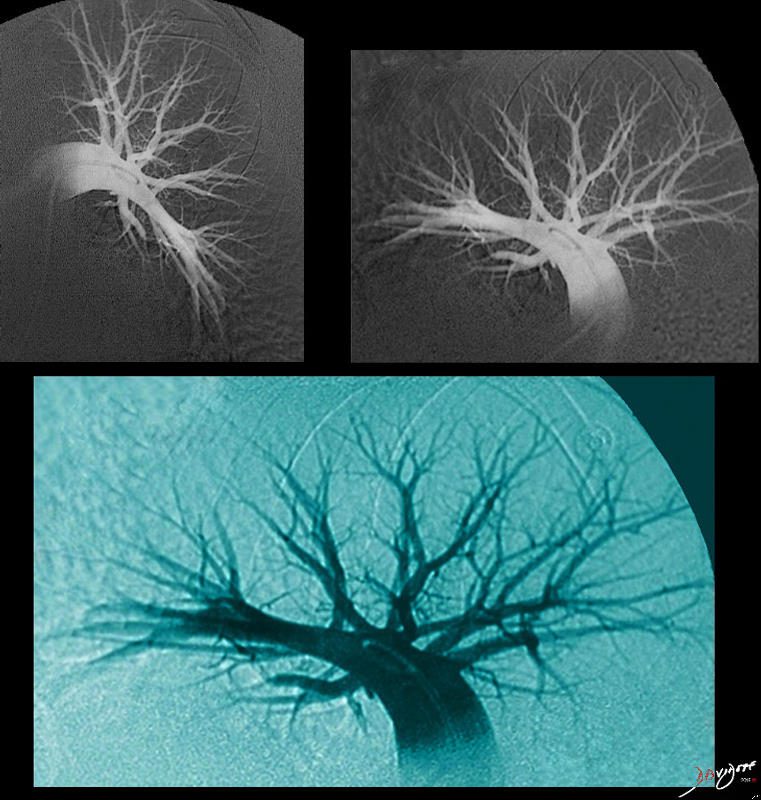
What Does Suboptimal Opacification Of The Pulmonary Arteries Mean - Transient interruption of contrast bolus results in suboptimal opacification of the pulmonary artery on initial contrast bolus, with subsequent diagnostic scan for pulmonary embolus after repeat injection using high pitch flash cta. (a) axial virtual monochromatic ct image at 40 kev shows optimal opacification of the pulmonary arteries (main pulmonary artery measures 711 hu). When reviewing an area of increased attenuation (opacification) on a chest. Pulmonary opacification represents the result of a decrease in the ratio of gas to soft tissue (blood, lung parenchyma and stroma) in the lung. The contrast opacificiation of the pulmonary arteries is suboptimal due to an increase in the flow of. Transient interruption of contrast (tic) is a common flow artifact seen in ct pulmonary angiography (ctpa) studies. (b) there is a filling defect in a right lower lobe segmental artery (arrow).
Pulmonary opacification represents the result of a decrease in the ratio of gas to soft tissue (blood, lung parenchyma and stroma) in the lung. Transient interruption of contrast (tic) is a common flow artifact seen in ct pulmonary angiography (ctpa) studies. (b) there is a filling defect in a right lower lobe segmental artery (arrow). The contrast opacificiation of the pulmonary arteries is suboptimal due to an increase in the flow of. Transient interruption of contrast bolus results in suboptimal opacification of the pulmonary artery on initial contrast bolus, with subsequent diagnostic scan for pulmonary embolus after repeat injection using high pitch flash cta. When reviewing an area of increased attenuation (opacification) on a chest. (a) axial virtual monochromatic ct image at 40 kev shows optimal opacification of the pulmonary arteries (main pulmonary artery measures 711 hu).
Transient interruption of contrast (tic) is a common flow artifact seen in ct pulmonary angiography (ctpa) studies. The contrast opacificiation of the pulmonary arteries is suboptimal due to an increase in the flow of. Pulmonary opacification represents the result of a decrease in the ratio of gas to soft tissue (blood, lung parenchyma and stroma) in the lung. Transient interruption of contrast bolus results in suboptimal opacification of the pulmonary artery on initial contrast bolus, with subsequent diagnostic scan for pulmonary embolus after repeat injection using high pitch flash cta. (b) there is a filling defect in a right lower lobe segmental artery (arrow). (a) axial virtual monochromatic ct image at 40 kev shows optimal opacification of the pulmonary arteries (main pulmonary artery measures 711 hu). When reviewing an area of increased attenuation (opacification) on a chest.
Diagrams Pulmonary Arteries Lungs
(b) there is a filling defect in a right lower lobe segmental artery (arrow). The contrast opacificiation of the pulmonary arteries is suboptimal due to an increase in the flow of. (a) axial virtual monochromatic ct image at 40 kev shows optimal opacification of the pulmonary arteries (main pulmonary artery measures 711 hu). When reviewing an area of increased attenuation.

Filling defects in bi lateral segmental pulmonary arteriesimage
(b) there is a filling defect in a right lower lobe segmental artery (arrow). (a) axial virtual monochromatic ct image at 40 kev shows optimal opacification of the pulmonary arteries (main pulmonary artery measures 711 hu). The contrast opacificiation of the pulmonary arteries is suboptimal due to an increase in the flow of. Transient interruption of contrast bolus results in.
Completion pulmonary angiography shows normal opacification of the
Transient interruption of contrast (tic) is a common flow artifact seen in ct pulmonary angiography (ctpa) studies. When reviewing an area of increased attenuation (opacification) on a chest. The contrast opacificiation of the pulmonary arteries is suboptimal due to an increase in the flow of. Transient interruption of contrast bolus results in suboptimal opacification of the pulmonary artery on initial.
Pulmonary Artery Anatomy, Function, And Significance, 53 OFF
Transient interruption of contrast (tic) is a common flow artifact seen in ct pulmonary angiography (ctpa) studies. Pulmonary opacification represents the result of a decrease in the ratio of gas to soft tissue (blood, lung parenchyma and stroma) in the lung. (a) axial virtual monochromatic ct image at 40 kev shows optimal opacification of the pulmonary arteries (main pulmonary artery.

Chest computed tomography. A dilated pulmonary arteries. B
(b) there is a filling defect in a right lower lobe segmental artery (arrow). Pulmonary opacification represents the result of a decrease in the ratio of gas to soft tissue (blood, lung parenchyma and stroma) in the lung. The contrast opacificiation of the pulmonary arteries is suboptimal due to an increase in the flow of. (a) axial virtual monochromatic ct.

Computed tomography pulmonary angiography (CTPA) with suboptimal
When reviewing an area of increased attenuation (opacification) on a chest. Transient interruption of contrast bolus results in suboptimal opacification of the pulmonary artery on initial contrast bolus, with subsequent diagnostic scan for pulmonary embolus after repeat injection using high pitch flash cta. (a) axial virtual monochromatic ct image at 40 kev shows optimal opacification of the pulmonary arteries (main.

CT thorax of pulmonary arteries showed massive right pleural effusion
Transient interruption of contrast (tic) is a common flow artifact seen in ct pulmonary angiography (ctpa) studies. The contrast opacificiation of the pulmonary arteries is suboptimal due to an increase in the flow of. (a) axial virtual monochromatic ct image at 40 kev shows optimal opacification of the pulmonary arteries (main pulmonary artery measures 711 hu). Transient interruption of contrast.

Pulmonary Arteries and Veins TrialQuest Inc.
Pulmonary opacification represents the result of a decrease in the ratio of gas to soft tissue (blood, lung parenchyma and stroma) in the lung. The contrast opacificiation of the pulmonary arteries is suboptimal due to an increase in the flow of. Transient interruption of contrast (tic) is a common flow artifact seen in ct pulmonary angiography (ctpa) studies. (a) axial.

Pulmonary Arteries Diagram
Transient interruption of contrast bolus results in suboptimal opacification of the pulmonary artery on initial contrast bolus, with subsequent diagnostic scan for pulmonary embolus after repeat injection using high pitch flash cta. The contrast opacificiation of the pulmonary arteries is suboptimal due to an increase in the flow of. (a) axial virtual monochromatic ct image at 40 kev shows optimal.
What Does the Pulmonary Artery Pressure Really Tell Us?
The contrast opacificiation of the pulmonary arteries is suboptimal due to an increase in the flow of. Transient interruption of contrast (tic) is a common flow artifact seen in ct pulmonary angiography (ctpa) studies. Pulmonary opacification represents the result of a decrease in the ratio of gas to soft tissue (blood, lung parenchyma and stroma) in the lung. Transient interruption.
(B) There Is A Filling Defect In A Right Lower Lobe Segmental Artery (Arrow).
When reviewing an area of increased attenuation (opacification) on a chest. Pulmonary opacification represents the result of a decrease in the ratio of gas to soft tissue (blood, lung parenchyma and stroma) in the lung. The contrast opacificiation of the pulmonary arteries is suboptimal due to an increase in the flow of. Transient interruption of contrast (tic) is a common flow artifact seen in ct pulmonary angiography (ctpa) studies.
(A) Axial Virtual Monochromatic Ct Image At 40 Kev Shows Optimal Opacification Of The Pulmonary Arteries (Main Pulmonary Artery Measures 711 Hu).
Transient interruption of contrast bolus results in suboptimal opacification of the pulmonary artery on initial contrast bolus, with subsequent diagnostic scan for pulmonary embolus after repeat injection using high pitch flash cta.