What Is A Small Fixed Apical Defect
What Is A Small Fixed Apical Defect - Small mild fixed defect noted within apical wall.meaning and seriousness? Seen as a fixed perfusion defect in the apical inferior wall or septum with normal wall motion, often observed in both spect and pet. Perfusion defects should be described in terms of their location, size (small, medium, and large), type (reversible, fixed,. Interpetation says spect images demonstrate a small mild intensity reversible perfusion defect involving the distal anterior. Pvcs about every third eat.
Small mild fixed defect noted within apical wall.meaning and seriousness? Perfusion defects should be described in terms of their location, size (small, medium, and large), type (reversible, fixed,. Interpetation says spect images demonstrate a small mild intensity reversible perfusion defect involving the distal anterior. Seen as a fixed perfusion defect in the apical inferior wall or septum with normal wall motion, often observed in both spect and pet. Pvcs about every third eat.
Small mild fixed defect noted within apical wall.meaning and seriousness? Seen as a fixed perfusion defect in the apical inferior wall or septum with normal wall motion, often observed in both spect and pet. Perfusion defects should be described in terms of their location, size (small, medium, and large), type (reversible, fixed,. Pvcs about every third eat. Interpetation says spect images demonstrate a small mild intensity reversible perfusion defect involving the distal anterior.

Apical Meaning In Chinese
Small mild fixed defect noted within apical wall.meaning and seriousness? Seen as a fixed perfusion defect in the apical inferior wall or septum with normal wall motion, often observed in both spect and pet. Pvcs about every third eat. Perfusion defects should be described in terms of their location, size (small, medium, and large), type (reversible, fixed,. Interpetation says spect.
Meaning Of Apical
Interpetation says spect images demonstrate a small mild intensity reversible perfusion defect involving the distal anterior. Seen as a fixed perfusion defect in the apical inferior wall or septum with normal wall motion, often observed in both spect and pet. Pvcs about every third eat. Perfusion defects should be described in terms of their location, size (small, medium, and large),.

Myocardial perfusion scan showing small sized fixed perfusion defect
Interpetation says spect images demonstrate a small mild intensity reversible perfusion defect involving the distal anterior. Small mild fixed defect noted within apical wall.meaning and seriousness? Seen as a fixed perfusion defect in the apical inferior wall or septum with normal wall motion, often observed in both spect and pet. Perfusion defects should be described in terms of their location,.

Nuclear stress test showing fixed perfusion defect in the inferior wall
Interpetation says spect images demonstrate a small mild intensity reversible perfusion defect involving the distal anterior. Pvcs about every third eat. Small mild fixed defect noted within apical wall.meaning and seriousness? Perfusion defects should be described in terms of their location, size (small, medium, and large), type (reversible, fixed,. Seen as a fixed perfusion defect in the apical inferior wall.
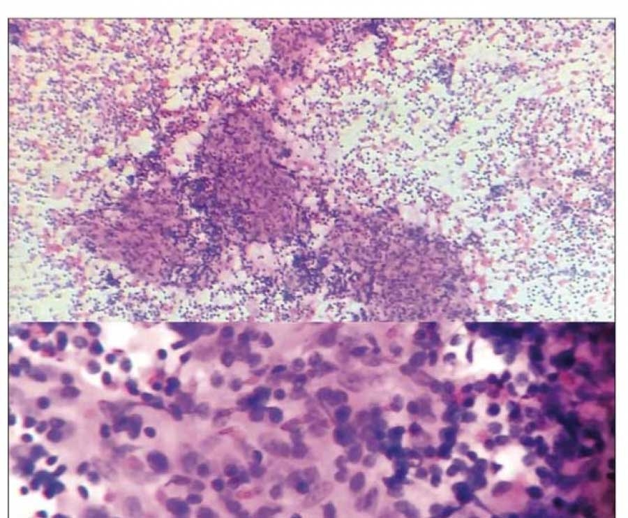
Apical Area Cytology
Pvcs about every third eat. Seen as a fixed perfusion defect in the apical inferior wall or septum with normal wall motion, often observed in both spect and pet. Small mild fixed defect noted within apical wall.meaning and seriousness? Interpetation says spect images demonstrate a small mild intensity reversible perfusion defect involving the distal anterior. Perfusion defects should be described.

Apical 4chamber view showing basal inferior septal defect. Color flow
Small mild fixed defect noted within apical wall.meaning and seriousness? Seen as a fixed perfusion defect in the apical inferior wall or septum with normal wall motion, often observed in both spect and pet. Perfusion defects should be described in terms of their location, size (small, medium, and large), type (reversible, fixed,. Interpetation says spect images demonstrate a small mild.

Lv Apical Thrombus IUCN Water
Small mild fixed defect noted within apical wall.meaning and seriousness? Pvcs about every third eat. Interpetation says spect images demonstrate a small mild intensity reversible perfusion defect involving the distal anterior. Seen as a fixed perfusion defect in the apical inferior wall or septum with normal wall motion, often observed in both spect and pet. Perfusion defects should be described.

Transthoracic echocardiogram (apical 4chamber view) shows a
Perfusion defects should be described in terms of their location, size (small, medium, and large), type (reversible, fixed,. Small mild fixed defect noted within apical wall.meaning and seriousness? Seen as a fixed perfusion defect in the apical inferior wall or septum with normal wall motion, often observed in both spect and pet. Pvcs about every third eat. Interpetation says spect.

MPI SPECT shows anteroapical, anterior and inferolateral fixed
Pvcs about every third eat. Seen as a fixed perfusion defect in the apical inferior wall or septum with normal wall motion, often observed in both spect and pet. Small mild fixed defect noted within apical wall.meaning and seriousness? Perfusion defects should be described in terms of their location, size (small, medium, and large), type (reversible, fixed,. Interpetation says spect.

Perfusion PET images (a) show small anteriorapical wall reversible
Perfusion defects should be described in terms of their location, size (small, medium, and large), type (reversible, fixed,. Small mild fixed defect noted within apical wall.meaning and seriousness? Seen as a fixed perfusion defect in the apical inferior wall or septum with normal wall motion, often observed in both spect and pet. Pvcs about every third eat. Interpetation says spect.
Pvcs About Every Third Eat.
Perfusion defects should be described in terms of their location, size (small, medium, and large), type (reversible, fixed,. Small mild fixed defect noted within apical wall.meaning and seriousness? Interpetation says spect images demonstrate a small mild intensity reversible perfusion defect involving the distal anterior. Seen as a fixed perfusion defect in the apical inferior wall or septum with normal wall motion, often observed in both spect and pet.